Finite Element Method

Interdepartmental Laboratory of Numerical Modeling

Finite Element Method

In the field of agricultural sciences, numerical modelling has proven to be a valuable tool in finding solutions to practical and scientific issues. Such models are necessary to understand the structure related properties of plants, like fruits and vegetables, and bring, in the next step, possibilities of extension by nano-structural features (like cell wall composition). This knowledge will be useful for engineering and improve fruits and vegetables quality.


The basic idea behind finite element method (FEM) is subdivision of a spatial domain of the problem into a simpler parts called the finite elements. The solution in global domain is obtained as a result of assembly of local solutions for the finite elements. Finite element method allows studies of systems with large complexity, irregular shapes and nonhomogeneous material properties.


Studies carried out by the Interdepartmental Laboratory of Numerical Modeling are aimed to create computational models of plant tissues at different spatial scales.

Modelling of plant tissues mechanical properties


Physical properties plant materials are related to several micro- and macroscopic morphological features such as the spatial arrangement and shape of cells, the number of intercellular spaces, turgor, the nano-composition of cell walls and the degree of degradation of the middle lamellae. The experimental analysis of the structure related micromechanical properties of plants  has been constrained by the lack of technology available for conducting reliable measurements at such scales. Deeper understanding of mechanical properties of plant tissues can be achieved by means of the numerical models of tissue deformation under various load conditions.


Up to now, the majority of models described in the literature was based on the principles of classical continuum mechanics. Most often, tissues were described by empirical or analytical models, as uniformly stressed structures, with symmetry and uniformity assumptions on cell shape and cell wall deformations. Although this approach allowed for fairly accurate predictions of the behavior of the plant materials in engineering applications, it was unable to provide an explanation of the micro-scale mechanisms underlying deformation and failure of tissue.


The research was co-founded by National Science Centre of Poland (research grant no. 2011/01/N/NZ9/02496).


Tissue structure modelling

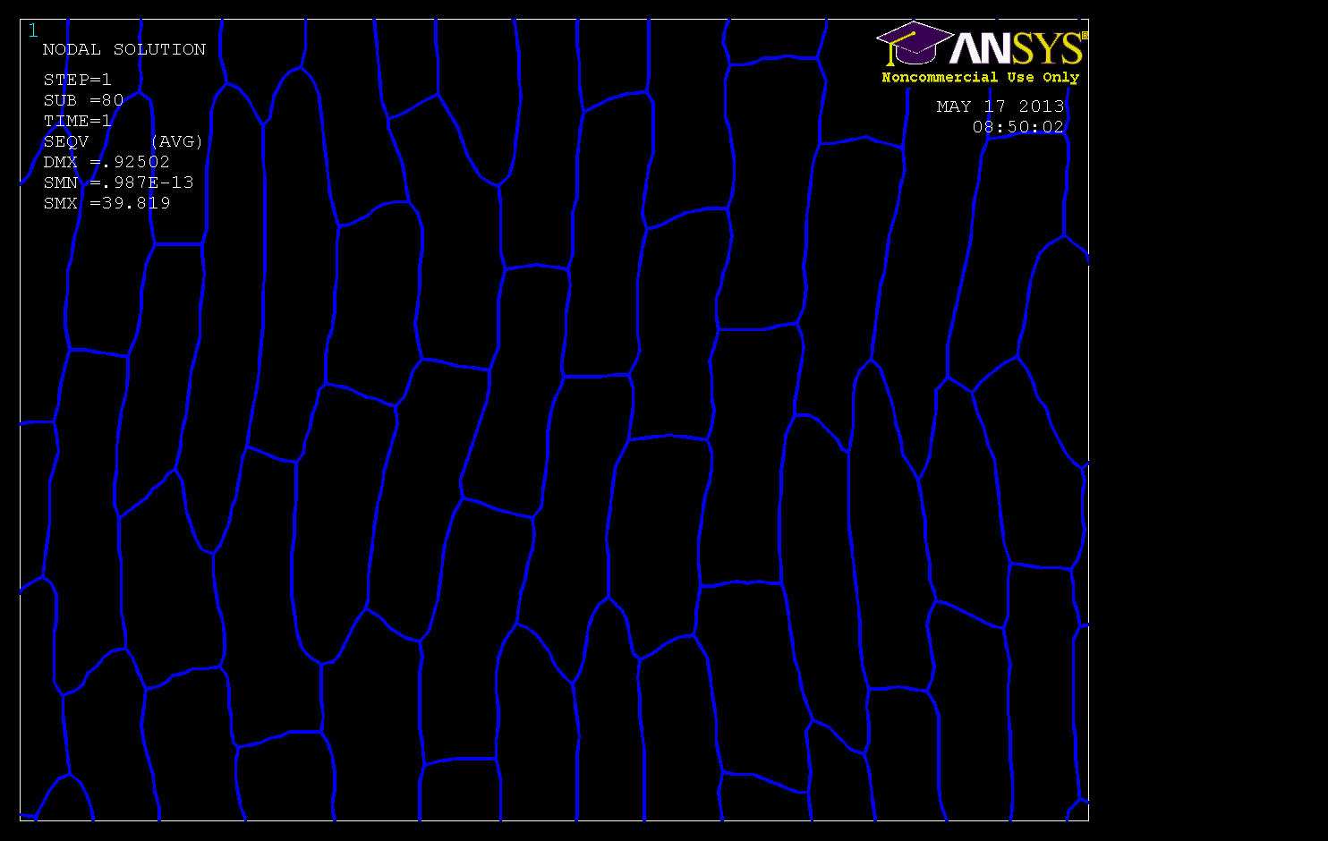
One of the important steps in solving a problem, before the appropriate analysis with FEM, is to create a virtual model of the tested object. The virtual model is defined by its geometry, material properties and boundary conditions such as loads and the type of supports. It is important to create a proper model with the highest possible degree of accuracy regarding real shape reconstruction; but on the other hand, the model must be simple enough to allow for efficient calculations.


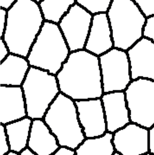
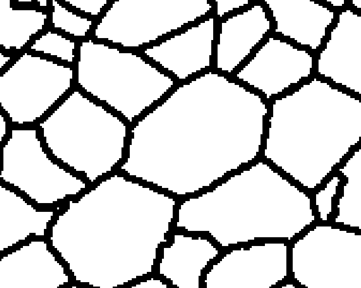
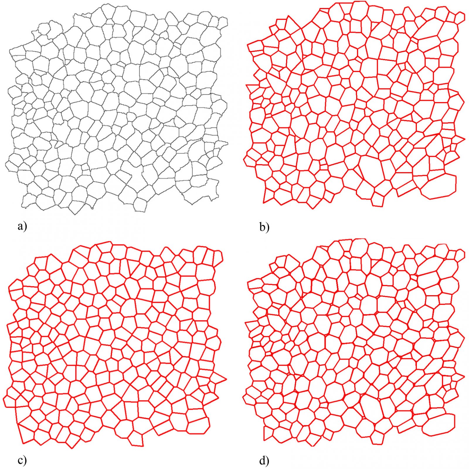
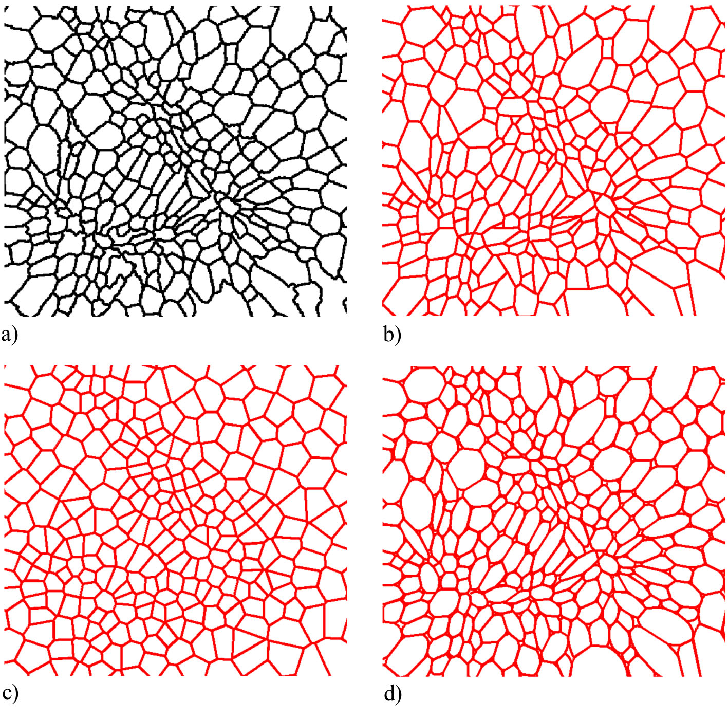
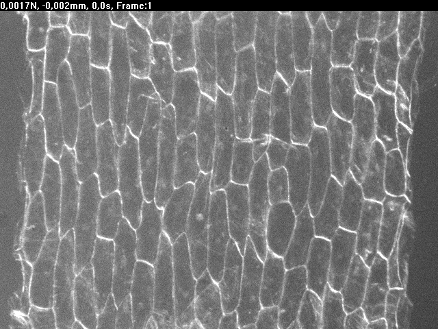
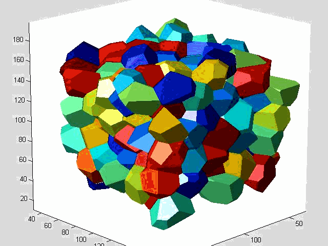
In the preliminary study three different methods for parameterisation of plant tissues were tested (Pieczywek et al., 2011).  All methods can be applied to images obtained with a confocal scanning laser microscope to create models for the simulation of the mechanical behaviour of biological cellular structures. Vectorisation, Voronoi tessellation and ellipse tessellation were tested. Potato tuber and carrot parenchyma were chosen as examples.

Badania finansowane były w ramach projektu nr 2011/01/N/NZ9/02496  Narodowego Centrum Nauki.

Modelowanie struktury tkanki


Jednym z kroków poprzedzających właściwą analizę MES jest utworzenie wirtualnej reprezentacji badanego obiektu. Wirtualny model zdefiniowany jest przez geometrię, właściwości materiałowe oraz warunki brzegowe reprezentujące oddziaływania zewnętrze oraz wewnętrzne. Istotne jest aby odwzorować modelowany system z możliwie największą dokładnością, przy jednoczesnym zachowaniu pewnych uroszczeń, umożliwiających przeprowadzenie obliczeń w rozsądnym czasie.


W badaniach wstępnych przetestowane zostały trzy różne metody parametryzacji struktury tkanki roślinnej (Pieczywek et al., 2011). Wszystkie metody zastosować można do obrazów mikroskopowych uzyskanych przy użyciu laserowego mikroskopu konfokalnego (CLSM), aby następnie utworzyć na ich podstawie wirtualne modele numeryczne struktur komórkowych. Testom poddano metodę wektoryzacji, teselacji eliptycznej, oraz teselacji Woronoja. Jako materiał badawczy wybrane zostały tkanki parenchymatyczne ziemniaka oraz marchwi.

Fig. 1. Vectorisation procedure: detection of a single cell boundary, detection of junction points,

connection of vertices and formation of a virtual cell.

Fig. 2. The process of creating a Voronoi diagram.

Fig. 3. The ellipse tessellation algorithm: fitting of ellipses, determination of the intersection points,

construction of “cutting lines”, the final model.

For each method tested, five geometrical parameters were analysed: area, perimeter, orientation, elongation and a local indicator of spatial association of all individual regions which represented cells. The reconstruction accuracy of the original tissue microstructure by each parameterisation method was investigated by the comparison of the geometrical properties of the cells from the segmentation with their virtual equivalents.

Fig. 4. Example of the parametrisation methods of the skeleton obtained from confocal scanning microscopy, a) original image of potato tissue, b) vectorisation, c) Voronoi tessellation, d) ellipse tessellation of the same structure.

Fig. 5. Performance of the parametrisation methods in reconstruction of irregularstructures: a) original image of carrot tissue, b) vectorisation, c) Voronoi tessellation, d) ellipse tessellation of the same structure.

Based on the results, Voronoi tessellation was considered to be inaccurate for tissue modelling. The vectorisation procedure only allowed for reproduction of the general shapes of cells, and the curvature of cell walls was neglected in this method. For both the Voronoi tessellation as well as vectorisation, created cells completely filled the space with no additional gaps and possessed sharp, angular shapes. The best overall reconstruction accuracy was obtained with ellipse tessellation. Models created with this method can be considered as representative equivalents of real tissues in terms of cell area, orientation, perimeter, shape and spatial arrangement.

Micro-scale model of onion epidermis tissue

In the present study a computational model of plant tissue that incorporates micro-scale geometrical features was developed to provide qualitative and quantitative predictions of the mechanical properties of onion (Allium cepa) epidermis (Pieczywek et al., 2014). Onion epidermal tissue has been chosen as a simple and well-defined system for model validation. The simulations of cellular structure behaviour under various mechanical load conditions were carried out using the finite element method (FEM).   The finite element method was chosen due to its computational efficiency, flexibility and ability to incorporate geometric nonlinearities. The models were validated against experimental data from a tensile test of the real tissue strips.

Fig. 6. Video record of tensile test using the real onion epidermis tissue.

Fig. 7. Animation showing tensile test using the FEM model of onion epidermis tissue.

The models showed capabilities of simulating large strains with nonlinear behaviour and produced force-strain curves that closely matched the experimental data. The model revealed a significant influence of tissue structure on micromechanical properties and allowed for interpretation of the tensile test results (force-strain curves) with respect to changes occurring in the structure of the virtual tissue. This proved that qualitative improvement of the results obtained from FEM models of plant tissues is possible due to incorporation of the real microstructure.

Fig. 8. Experimental stress-strain curve for tensile test of onion epidermis tissue. E1 – elasticity modulus in the first part of the curve, E2 – modulus of elasticity in second linear part of curve (after transition to elasto-plastic deformation), σpl and εpl – stress and strain at the beginning of the transition phase, respectively.

Fig. 9. FEM models performance for three cell wall parameters: E1exp, E1mod – elasticity modulus in the first part of the curve, E2exp, E2mod – elastic modulus in second linear part of curve, σpl exp, σpl mod – stress and at the yielding point.

Presented model shows innovative approach to modelling of plant micromechanics and the efforts undertaken so far in this field were performed only by a few research institutions worldwide. Although the model has been developed on a relatively simple epidermal tissue, it can be generalized to more complicated tissue with a different shapes of cells and intercellular spaces. Such models are necessary to understand the structure related properties of plants, like fruits and vegetables, and bring, in the next step, possibilities of extension by nano-structural features (like cell wall composition). This knowledge will be useful for engineering and improve fruits and vegetables quality, for instance by growing varieties of fruit, which, due to the morphological characteristics of tissue would be more resistant to postharvest damage or would be developed for specific consumer preferences.

Future work


Simulations of fully 3D, automatic generated cellular structures.

X