Department of Microstructure and Mechanics of Biomaterials

Equipment

Confocal Laser Scanning Microscope (CLSM) – FluoView300


  • CLSM allows for micro-structure observation of samples surfaces and 3D imaging. Images are taken only from thin confocal layer, thus images are usually very contrastic.
  • Main features: Two lasers: Ar 488 nm,  HeNe 543 nm, 3 channels (two confocal, one optical), high resolution; XY, XYZ, XYZT scanning modes.

Atomic Force Microscope (AFM) – BIOSCOPE CATALYST II


  • AFM allows for nano- and micro-structure observation with analysis of mechanical properties at these scales in air or liquid of biological samples.
  •  Main features: Contact, tapping, force volume modes; Harmonix mode for mechanical properties imaging; system for perfusion; temperature control, integration of optical and AFM view .

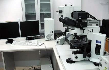
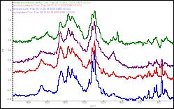
Fourier-transform infra-red spectroscopy (FTIR)/Raman spectroscopy system


The system includes Raman disperse spectrometer with 532 nm laser, Fourier-transform infra-red Raman spectrometer
with 1064 nm laser, IR spectrometer and UV-VIS spectrometer.

  • Structural identification and analysis of chemical compounds, mapping of chemical compound distribution in samples.

Examples of use:

  • Analysis of multi-molecular compounds; analysis of cellulose crystallinity.
  • Distribution of active substances in pharmaceutical tablets; chemometric polymer analysis.

Aphelion Dev. 4.0 image analysis software


  • Computer analysis of 2D and 3D images.
  • 2D and 3D image transformation, and analyses, including colour, size and orientation of represented objects.

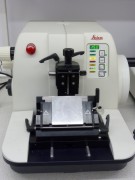
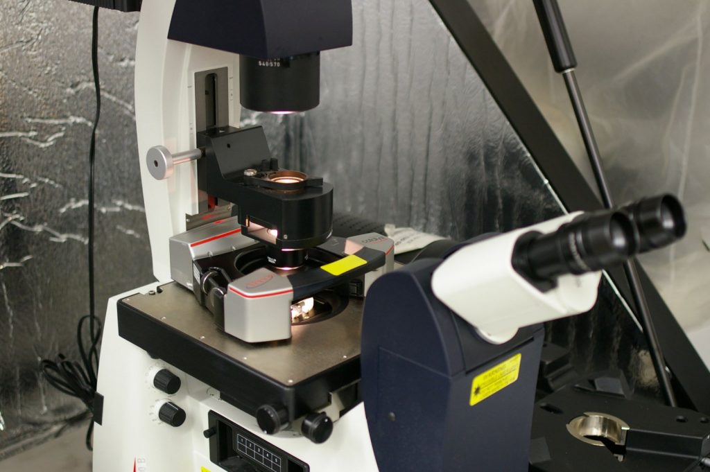
Microtome – LEICA RM2155


Microtome is a tool for slicing fixed samples with high precision (< 1 micrometer).

Main features: Cutting accuracy <1 micron, fully programmable; possibility to use different blades: glass, stainless steel, sapphire.


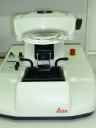
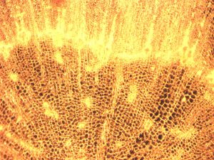
Vibratome – LEICA VT 1000S


Vibratome is useful for slicing soft materials like plant and animal tissues. Surface has decent quality for CLSM or macroscope.

Main features: Adjustable thickness (with 1 micron step), cutting speed and oscillations; batch for sample allowing cutting in buffers; fully programmable; possibility of use simple razors blades.


Macroscope


  • Macroscope is a microscope using the dark field view. It is designed for observation large sample areas. Sample must be transparent.
  • Main feature: field of view up to~8x6mm. Useful for evaluation of samples heterogeneity.
X